
A Case of Primary Intracranial Angiomyoma and Review of the Literature
##plugins.themes.bootstrap3.article.sidebar##
##plugins.themes.bootstrap3.article.main##
Abstract
Background. A rare presentation of angiomyoma (also called angioleiomyoma or ALM) is primary intracranial ALM (ICALM). This benign tumor is more common in men aged 40-60 years but its etiology remains unclear. Because of limited reports, atypical clinical manifestations and imaging features, ICALM is often misdiagnosed.
Methods. We describe the case of a 58-year-old male ICALM patient admitted with intermittent left side headaches and conduct a literature review to summarize the key clinical, imaging and pathological features of all known cases of ICALM.
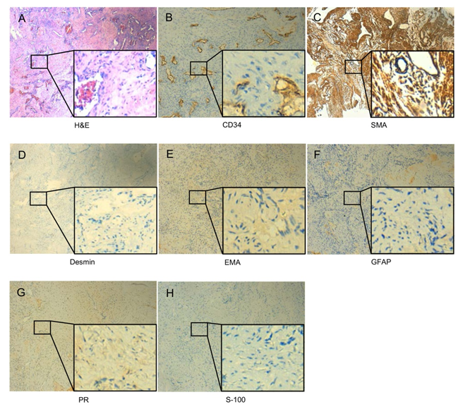
Results. Only 53 known cases of ICALM have been previously reported and we describe the first case instance located in the left cerebellopontine angle area. This patient's preoperative magnetic resonance imaging (MRI) imaging showed homogeneously T1-weighted low signal,slightly T2-weighted high signal and the diffusion weighted imaging (DWI) slightly low signal, which was consistent with the MRI features of known cases of ICALM. The patient underwent tumor resection through the right retrosigmoid sinus approach with an uneventful postoperative course and no long-term complications. Pathological diagnosis confirmed ICALM with tumor cells positive for smooth muscle actin and negative for neurological, mesenchymal and epithelial markers. Analysis of the literature showed that the cavernous type of ICALM was most common with our case example being a mixture of solid and cavernous types.
Conclusions. The progressive enhancement of MRI can contribute to ICALM diagnosis with pathological confirmation by immunohistochemistry. The first-line treatment strategy for ICALM is surgical resection, which can lead to good outcomes.