
Differentially Expressed Genes and Their Signaling Pathways in ARPE-19 Cells Induced by 448 nm Blue Light
##plugins.themes.bootstrap3.article.sidebar##
##plugins.themes.bootstrap3.article.main##
Abstract
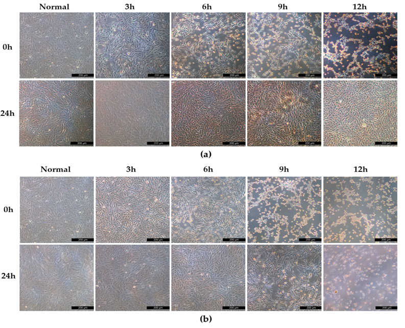
The enduring exposure to blue light emerges as a potential contributor to retinal degenerative diseases, with damage to the retinal pigment epithelium identified as the primary source of light-induced retinal damage. This study delves into the detrimental effects of 448 nm blue light on human retinal pigment epithelial (ARPE-19) cells and the underlying mechanisms. ARPE-19 cells underwent irradiation with 25 mW/cm² of blue light for durations ranging from 3 to 12 hours. Comprehensive analyses, including transcriptome sequencing and RT-qPCR, were employed to assess alterations across different temporal groups. Following blue light exposure, the expression profile of ARPE-19 cells underwent significant changes. In the 6-hour group compared to the normal group, a total of 4894 differentially expressed genes (DEGs) were identified. Transcriptome sequencing analysis revealed an upregulation of apoptosis-related genes after 6-hours of blue light exposure. Correspondingly, RT-qPCR detection demonstrated that blue light could induce ferroptosis in ARPE-19 cells. Specifically, the expression levels of ferroptosis-related genes, including NCOA4, FTH1, FTL, SLC39A14, LPCAT3, HMOX1, SLC7A11, and P53, exhibited a significant increase, while the expression levels of SLC3A2 and GPX4 were significantly reduced. Blue light irradiation induced notable alterations in the cell expression profile, with ferroptosis occurring at the early stages of irradiation. The findings from this study provide valuable insights for the safe use of visible light laser. The paper should interest readers in the areas of laser-induced ocular damage, laser dazzling effects, and the development of laser dazzling devices.